Sim Cliff Notes - SEPTEMBER 2017
/Every month we summarize our simulation cases. No deep dive here, just the top 5 takeaways from each case. This month's cases included anaphylaxis, PCP pneumonia, neutropenic fever, Giant Cell arteritis and Polymalgia Rheumatica.
5 Step approach to Anaphylaxis
1. Identify Anaphylaxis
- It can sometimes be difficult to recognize anaphylaxis because it can mimic other conditions and be variable in its presentation.
- Because of the wide spectrum of signs and symptoms, the diagnostic criteria can be hard to remember.
- I like to think of anaphylaxis as a sudden onset, systemic allergic reaction that includes either wheezing, hypotension, or oropharyngeal edema and requires the immediate administration of epinephrine.
- It is important to note that only 8/10 have skin findings.
2. Give epinephrine early
- Epinephrine is the first line treatment.
- #1 cause of death is failure to give epinephrine early.
- The dose 0.3-0.5 mg 1:1,000 IM, 0.01 mg/kg in children (max 0.3 mg); administer every 5 min x 3.
- For effectiveness: intramuscular trumps subcutaneous, the thigh muscle trumps the deltoid.
3. Give supporting medications
- Benadryl – blocks antihistamine but does not work alone
- H2 blocker – blocks bradykinin (causes angioedema, urticaria)
- Steroids - block potential rebound anaphylaxis
- IV fluids - improve hypotension in vasodilatory state
- Consider glucagon for patients on beta blockers and little improvement with IM epinephrine
4. Consider IV epinephrine if your patient is not responding to several doses of IM epinephrine
- Take the “crash cart” epi (1 mg (1:10,000) epinephrine in 10 ml) and put in a 1000 ml NS bag
- You now have an epinephrine drip at 1 mcg/ml
- Start at 1cc (1 ml)/min into a high flow IV
- Titrate to effect in 1-3 min (2-10 mcg/min)
Watch Dr. Corey Slovis discuss how to make an epinephrine drip
5. Be aware of rebound anaphylaxis
- Past studies have quoted an incidence of 5-20% but new literature has challenged this
- It is reasonable to observe patients who have responded to treatment for anaphylxis for at least 6 hours
- Make sure they are discharged with an epinephrine auto-injector
References
1. Lieberman P, Nicklas RA, Randolph C, et al. American Academy of Allergy, Asthma, and Immunology/American College of Allergy, Asthma, and Immunology (AAAAI/ACAAI) Joint Task Force: Anaphylaxis - a practice parameter update 2015. Ann Allergy Asthma Immunol. 2015 Nov;115(5):341-84.
2. Sampson HA, Muñoz-Furlong A, Campbell RL, et al. Second symposium on the definition and management of anaphylaxis: summary report Second National Institute of Allergy and Infectious Disease/Food Allergy and Anaphylaxis Network symposium. J Allergy Clin Immunol. 2006 Feb;117(2):391-7.
3. Simons FE. Anaphylaxis. J Allergy Clin Immunol. 2010 Feb;125(2 Suppl 2):S161-81, correction can be found in J Allergy Clin Immunol 2010 Oct;126(4):885.
4. National Institute of Health and Care Excellence. Anaphylaxis: assessment to confirm an anaphylactic episode and the decision to refer after emergency treatment for a suspected anaphylactic episode. NICE 2011 Dec:CG134 PDF, executive summary can be found in BMJ 2011 Dec 14;343:d7595.
5. Campbell RL, Li JT, Nicklas RA, Sadosty AT, Members of the Joint Task Force, Practice Parameter Work Group. Emergency department diagnosis and treatment of anaphylaxis: a practice parameter. Ann Allergy Asthma Immunol. 2014 Dec;113(6):599-608.
6. Ellis AK, Day JH. Incidence and characteristics of biphasic anaphylaxis: a prospective evaluation of 103 patients. Ann Allergy Asthma Immunol. 2007;98:64-69.
7. Smit DV, Cameron PA, Rainer TH. Anaphylaxis presentations to an emergency department in Hong Kong: incidence and predictors of biphasic reactions. J Emerg Med. 2005;28:381-388.
8. Pumphrey RSH. Lessons for management of anaphylaxis from a study of fatal reactions. Clin Exp Allergy. 2000;30:1144-1150. (Retrospective; 164 patients)
9. Lee S, Bellolio MF, Hess EP, Erwin P, Murad MH, Campbell RL. Time of Onset and Predictors of Biphasic Anaphylactic Reactions: A Systematic Review and Meta-analysis. J Allergy Clin Immunol Pract. 2015 May-Jun;3(3):408-16.e1-2. Epub 2015 Feb 11.
10. Grunau, BE. Incidence of Clinically Important Biphasic Reactions in Emergency Department Patients With Allergic Reactions or Anaphylaxis Annals of Emerg Med 2014;63:736-744.
HIV/AIDS patients with respiratory infections
1. As with all HIV patients, put their clinical picture in the context of their last CD4 count.
- A CD4 count less than 200/mm3 leads to more advanced disease and makes HIV patients more susceptible to opportunistic infections.
- Patients with a CD4 count > 200/mm3 are basically immuno-competent and can be treated like anyone else.
2. Mycobacterium tuberculosis (TB) should be considered in all HIV-infected patients with pneumonia, and it often presents atypically in these patients. These patients should be placed in isolation until TB can be ruled out.
3. Radiographic findings of pneumonia are highly variable in HIV disease.
- Classic findings for the three major categories of HIV – related pneumonias (P. jiroveci, community – acquired bacterial pneumonia and TB) may be absent.
- PCP cannot be reliably distinguished from bacterial pneumonia or TB based on symptoms or xray findings.
There are diffuse bilaterally symmetric interstitial patten noted in the perihilar region and extending towards the periphery. Multiple ill defined small hyperlucent patches are noted in the bilateral lung fields especially in the mid zones suggestive of pneumatocele.
https://radiopaedia.org/cases/pneumocystis-carinii-pneumonia-1
4. While there may be clues in the patient’s presentation and laboratory results that suggest a particular etiology, one safe approach is to address all of the most common pathogens (P. jiroveci, community acquired bacterial pneumonia, and TB) in each patient.
5. Patients who have PCP pneumonia and hypoxia (<90%) have a reduced mortality if steroids are administered.
References
1. Sokolove PE, Rossman L, Cohen SH. The emergency department presentation of patients with active
pulmonary tuberculosis. Acad Emerg Med 2000 Sep;7(9):1056-1060.
2. Moran GJ, McCabe F, Morgan MT, et al. Delayed recognition and infection control for tuberculosis
patients in the emergency department. Ann Emerg Med 1995 Sep;26(3):290-295.
3. Hopewell PC. Pneumocystis carinii pneumonia: diagnosis. J Infect Dis 1988 Jun;157(6): 1115-1119.
4. Magnenat JL, Nicod LP, Auckenthaler R, et al. Mode of presentation and diagnosis of bacterial pneumonia
in human immunodeficiency virus-infected patients. Am Rev Respir Dis 1991 Oct;144(4):917-922.
5. Havlir DV, Barnes PF. Tuberculosis in patients with human immunodeficiency virus infection. N Engl J
Med 1999 Feb 4;340(5):367-373.
6. Ewald H, Raatz H, Boscacci R, Furrer H, Bucher HC, Briel M. Adjunctive corticosteroids for Pneumocystis jiroveci pneumonia in patients with HIV infection. Cochrane Database Syst Rev. 2015.
7. Montaner JS, Lawson LM, Levitt N, Belzberg A, Schechter MT, Ruedy J Corticosteroids prevent early deterioration in patients with moderately severe Pneumocystis carinii pneumonia and the acquired immunodeficiency syndrome (AIDS). Ann Intern Med. 1990;113(1):14.
8. Bozzette SA, Sattler FR, Chiu J, Wu AW, Gluckstein D, Kemper C, Bartok A, Niosi J, Abramson I, Coffman J. A controlled trial of early adjunctive treatment with corticosteroids for Pneumocystis carinii pneumonia in the acquired immunodeficiency syndrome. California Collaborative Treatment Group. N Engl J Med. 1990;323(21):1451
5 step approach to Neutropenic Fever
1. Know the definition of febrile neutropenia.
- A single temperature measurement ≥ 38.3°C (101°F) or a sustained temperature ≥ 38°C (≥ 100.4°F) for more than 1 hour.
- ANC of either < 500 cells/μL or < 1000 cells/μL, with a predicted nadir of < 500 cells/μL over the subsequent 48 hours.
2. Treat febrile neutropenia promptly, aggressively and early to reduce mortality. Treat early with broad-spectrum bactericidal coverage against flora from gut, skin, and respiratory tract with third or fourth generation cephalosporin +/- gentomycin +/- vancomycin.
3. Perform a thorough history and physical exam looking for a source (including sometimes neglected areas like the mouth and perineum).
4. Consider the use of CT for highly suspicious findings on history/physical exam(eg. Sinus CT for sinus tenderness/purulent nasal discharge, chest CT for suspected pneumomia, or abdominal CT for diarrhea and abdominal pain).
5. Consider the use of Vancomycin if specific indications exist.
- Hypotension
- Preliminary cultures showing gram-positive flora
- Known history of methicillin-resistant Staphylococcus aureus or Β-lactam-resistant pneumococci
- Prior prophylaxis with a fluoroquinolone or trimethoprim/sulfamethoxazole
- Probable catheter-related infection
IDSA Clinical Practice Guideline for Neutropenic Fever
Freifeld AG, Bow EJ, Sepkowitz KA, et al. Clinical practice guideline for the use of antimicrobial agents in neutropenic patients with cancer: 2010 update by the infectious diseases society of america. Clin Infect Dis 2011; 52:e56.
Polymalgia rheumatica and
giant cell arteritis
1. What is Polymyalgia Rheumatica (PMR)?
- PMR is a common chronic, inflammatory rheumatic disease characterized by muscle pain and stiffness, particularly affecting the neck, shoulders, and hips.
- Contrary to the name, it is not a myopathic process. Proximal articular and periarticular structures are affected (joints, bursae, tendons), classically at the hips and shoulders.
2. Why should the emergency physician care about diagnosing it?
- It is relatively common, but often missed.
- Missed PMR is a cause of significant disability (and increased risk for falls).
- Up to 20% of patients with PMR have associated Giant Cell Arteritis (GCA), a potentially vision threatening illness.
- Patients with PMR have increased risk of stroke, MI.
- PMR is very responsive to steroids, a treatment most emergency physicians are comfortable initiating.
3. How do you make the diagnosis of PMR?
- There is no universally accepted diagnostic criteria.
- PMR should be suspected in patients over 50 with symptoms involving aching and stiffness about the upper arms, posterior neck, pelvic girdle, and/or lumbar region (all worse upon arising in morning).
- High yield questions to ask your patient include:
- “Have you had trouble putting on a shirt or coat? Trouble hooking a bra in the back?”
- “Do you have trouble standing due to pain?”
- “Have you required assistance with morning dressing?”
- "Gelling," or stiffness with inactivity, is a hallmark of synovitis in the systemic rheumatic diseases in general, but in PMR this phenomenon can be notably severe.
- In suspected cases, both ESR and CRP should be ordered (ESR can be < 40 mm/hour in up to 20% of patients).
4. Evaluate for Giant Cell Arteritis (GCA) in patients with suspected PMR
- GCA is a syndrome of systemic vasculitis commonly affecting the branches of the internal and carotid arteries (most common form of systemic vasculitis affecting patients > 50 years old).
- Involvement of these arterial branches leads to clinical findings of headache, jaw claudication, scalp tenderness and blindness.
- 40% - 60% of patients with GCA have PMR symptoms at diagnosis.
- 16% - 21% of patients with PMR have GCA.
5. So what is the emergency physician to do about it?
- Treatment of both PMR and GCA involves the initiation of steroids.
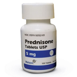
- PMR – prednisone 10-20 mg/day
- GCA – prednisone 40-60 mg/day
- Patients with suspected GCA should be referred for a temporal artery biopsy.
- There should be no delay in starting therapy because the artery biopsy will still show inflammatory changes for several days after corticosteroids have been started and the result is unlikely to alter therapeutic decisions.
References
1. Kermani TA, Warrington KJ. Polymyalgia rheumatica. Lancet. 2013 Jan 5;381(9860):63-72, correction can be found in Lancet. 2013 Jan 5;381(9860):28.
2. Dasgupta B, Borg FA, Hassan N, et al; British Society of Rheumatology and British Health Professionals in Rheumatology Standards, Guidelines and Audit Working Group. BSR and BHPR guidelines for the management of polymyalgia rheumatica. Rheumatology (Oxford). 2010 Jan;49(1):186-90.
3. Dejaco C, Singh YP, Perel P, et al. European League Against Rheumatism., American College of Rheumatology. 2015 recommendations for the management of polymyalgia rheumatica: a European League Against Rheumatism/American College of Rheumatology collaborative initiative. Arthritis Rheumatol. 2015 Oct;67(10):2569-80.
Written by Jeffrey A Holmes, MD


