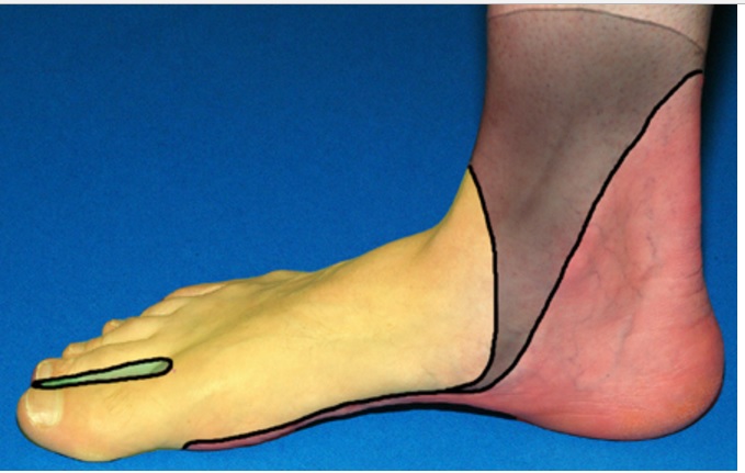


Take Note:
The superficial peroneal, sural, and saphenous nerves are located in the subcutaneous tissue encircling the ankle. These nerves branch out and anastomose extensively; therefore they do not have a single point that can be consistently anesthesitized. A field block in the subcutaneous tissue is used to anesthetize these nerves.
Deep Peroneal Nerve Block
Anatomy:
- The deep peroneal nerve lies in the groove between the extensor hallucis longus and the tibialis anterior tendon.
- The hallucis longus can be located by having the patient flex and extend the big toe.
- The tibialis interior can be located by having the patient dorsi flex the foot and invert the ankle.
- The injection site should be at the level of the superior malleolus and between the two tendons.
Distribution of anesthesia:
The deep peroneal nerve provides sensation to the web space between the first and second toe and a small area just proximal to the first and second toe on the plantar aspect of the foot.

Technique:
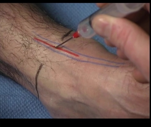
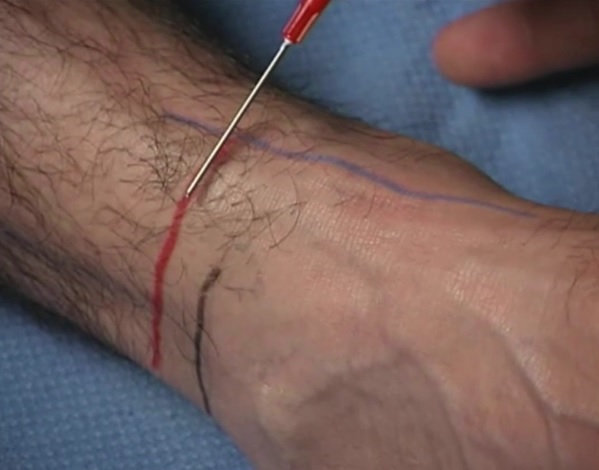
BLack = superior border of medial and lateral malleoli; blue = tibialis anterior and extensor hallicus longus; red = deep peroneal nerve
The surgical field should be prepared across the anterior surface of the ankle between the superior aspect of the medial and lateral malleoli. Raise a wheal of anesthesia in the subcutaneous space and direct the needle between the tendons of the hallucis longus and the tibialis anterior at the level of the superior malleoli. Insert the needle until it is deep to the tendons or bone is struck. Inject approximately 5 milliliters of anesthetic. Withdraw the needle and redirect thirty degrees laterally and then thirty degrees medially and provide an additional 3 to 5 ml of anesthetic. If anesthesia in the saphenous distribution is also desired, bring the needle back to the level of the subcutaneous tissue and redirect it medially towards the medial malleolus. Inject an additional 5 ml in the subcutaneous space. This will block the saphenous nerve.
Pitfalls:
- Avoid inadvertent saphenous vein puncture
- Intraneural injection will cause significant pain, therefore withdraw the needle a few millimeters and continue injecting anesthetic
Posterior Tibial Nerve Block

View from bottom of foot
Anatomy:
The posterior tibial nerve runs just behind the medial malleolus, and just posterior to the posterior tibial artery. Like the deep peroneal, the posterior tibial nerve is deep to the fascia. The posterior tibial nerve can be located just posterior to the medial malleolus just superficial to the artery.
Distribution of anesthesia:

Blue = achilles tendon; black = medial malleolus; red = Posterior tibial nerve
The posterior tibial nerve provides the majority of the sensation to the plantar aspect of the foot with minor contributions from the deep peroneal and sural nerve. The posterior tibial nerve also provides sensation to the heel of the foot. Technique: The surgical field should be prepared posterior to the medial malleolus. Identify the posterior tibial artery by palpating the artery posterior to the medial malleolus. Insert the needle just posterior to the artery until it penetrates the deep fascia. If the pop of the deep fascia cannot be felt, continue inserting the needle until it contacts bone. Withdraw the needle 2 to 5 millimeters and inject 3-5 5 milliliters of anesthesia. To increase the odds of a successful block, place an additional 3 to 5 milliliters lateral and medial to the original injection site.
Pitfalls:
Intraneural injection will cause excruciating pain with injection, therefore withdraw the needle a few millimeters and continue injecting the anesthetic.
Superficial Peroneal Nerve block
Anatomy:

The superficial peroneal nerve is superficial and runs along the anterior lateral portion of the ankle. It can be blocked by subcutaneous injection between the lateral malleolus and the tibialis anterior tendon.
Distribution of anesthesia:
black = superior portion of lateral malleolus; blue = tibialis anterior tendon; red = approximate injection point for superficial peroneal nerve
This nerve provides sensation to the dorsal lateral aspect of the foot. Technique: Identify the tibialis anterior tendon by having the patient dorsiflex the foot and inverts the ankle. The most prominent tendon should be the tibialis anterior. The surgical field should be prepared between the tibialis anterior tendon and the lateral malleolus at the level of the superior malleoli. Inject anesthesia in the subcutaneous space from the tibialis anterior tendon to the superior portion of the lateral malleolus.
Pitfalls:
Intraneural injection will cause significant pain, therefore withdraw the needle a few millimeters and continue injecting the anesthetic.
Sural Nerve
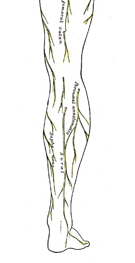

Black = Lateral malleolus; blue= achilles tendon; red = injection area
Anatomy:
The sural nerve is quite superficial and can be blocked by anesthetizing the subcutaneous tissue from the superior portion of the lateral malleolus to the Achilles tendon.
Distribution of anesthesia:
The sural nerve provides sensation to the lateral aspect of the ankle and a small area on the plantar lateral aspect of the foot.
Technique:
The surgical field should be prepared between the Achilles tendon and the lateral malleolus at the level of the superior malleoli. Inject anesthesia in the subcutaneous space from the superior portion of the lateral malleolus to the Achilles tendon.
Pitfalls:
Intraneural injection will cause significant pain, therefore withdraw the needle a few millimeters and continue injecting the anesthetic.
Saphenous Nerve

Anatomy:
The saphenous nerve is a subcutaneous nerve that can be blocked by injecting anesthesia from the superior medial malleolus to the tibialis anterior tendon. Use caution around the saphenous vein.
Distribution of anesthesia:
This nerve provides sensation to the medial aspect of the ankle.
Technique:

Black = medial malleolus; blue = tibialis anterior; red = approximate injection point
Identify the tibialis anterior tendon and the superior portion of the medial malleolus. The surgical field should be prepared between the tibialis anterior tendon and the medial malleolus at the level of the superior malleoli. Inject anesthesia in the subcutaneous space from the tibialis anterior tendon to the superior portion of the medial malleolus.
Pitfalls:
Puncture of the saphenous vein.
Intraneural injection will cause significant pain, therefore withdraw the needle a few millimeters and continue injecting the anesthetic.
Written by Douglas Dillon, MD
Edited and Posted by Jeffrey A Holmes, MD